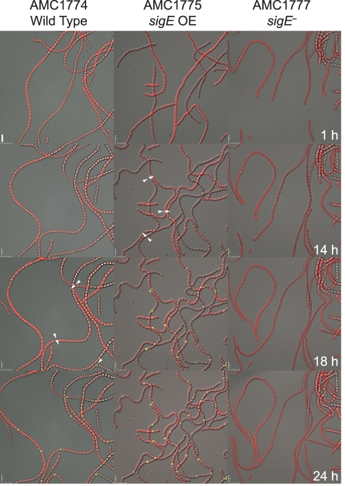
Fig. 5.
Time-lapse microscopy images of a nifHD-gfp reporter construct integrated into the nifH locus on the chromosome in wild-type (AMC1774), sigE overexpression (OE) (AMC1775), and sigE mutant (AMC1777) backgrounds. Images are shown for 1, 14, 18, and 24 h after nitrogen step-down. Images are merged DIC (grayscale), autofluorescence (red), and GFP reporter fluorescence (green). Arrows indicate weak GFP fluorescence from proheterocysts. Scale bars, 10 μm.

