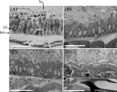
Fig. 7.
Histopathological effects of TC on the anterior midgut of P. xylostella epithelium. (A) Untreated larva, showing a regular arrangement of basal membrane (bm) and columnar cells (cc) in the midgut epithelium and the lumen (lu). (B to D) Larvae at 16 h (B), 24 h (C), and 40 h (D) after the larval ingestion of food dosed with 100 ng TC/cm2. Within 16 h, apical swelling of the columnar cells occurs with vesicle-like structures seen in the gut lumen. At 40 h, there is a complete dissolution of the larval gut lining and the presence of cellular debris in the lumen. Bars = 50 μm.

