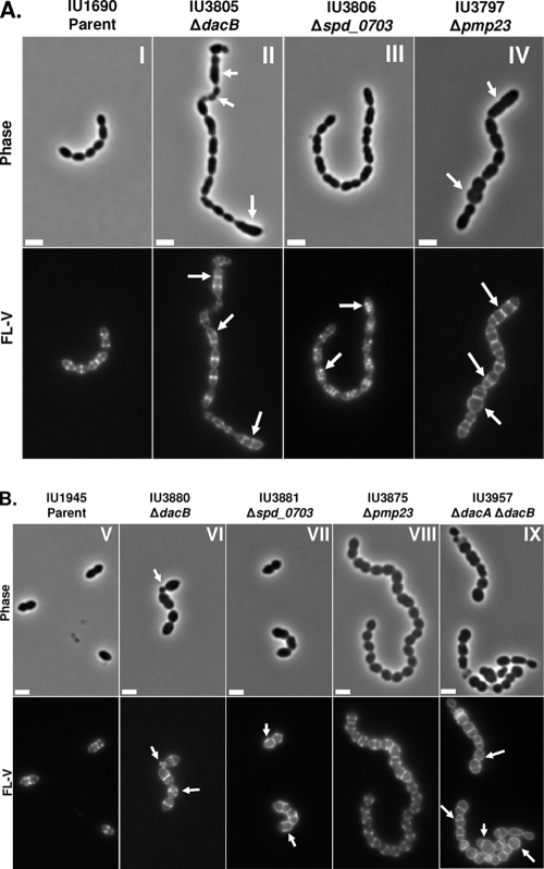
Fig. 2.
Representative phase-contrast micrographs (top rows) and FL-V staining (bottom rows) of deletion mutants lacking PG hydrolases or WalRKSpn regulon members. Labeling and microscopy were carried out numerous times using independent cultures as described in Materials and Methods. (A) Deletion mutants in encapsulated strain D39: panel I, parent (IU1690); panel II, ΔdacB (IU3805); panel III, Δspd_0703 (IU3806); panel IV, Δpmp23 (IU3797). (B) Deletion mutants in isogenic unencapsulated strain D39: panel V, D39 Δcps parent (IU1945); panel VI, ΔdacB (IU3880); panel VII, Δspd_0703 (IU3881); panel VIII, Δpmp23 (IU3875); panel IX, ΔdacA ΔdacB (IU3957). Arrows indicate defects in cell morphology (phase micrographs) or PG pentapeptide localization (FL-V micrographs) compared to the IU1690 or IU1945 parent strain. The scale bars correspond to 2 μm.

