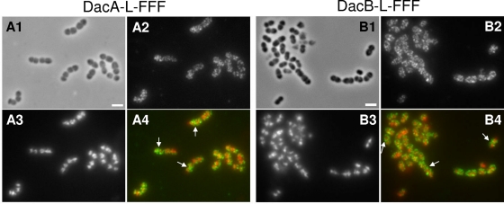
Fig. 6.
IFM localization of DacASpn-L-FFF and DacBSpn-L-FFF proteins in unencapsulated D39 pneumococcal cells growing exponentially in BHI broth. Strains IU4960 (A) and IU4961 (B) expressed DacASpn-L-FFF and DacBSpn-L-FFF, respectively, from their native chromosomal loci, where L is a linker segment and FFF is three tandem copies of the FLAG epitope tag (see Materials and Methods; Table 2) (see reference 69). A1 and B1, phase-contrast micrographs; A2 and B2, IFM using polyclonal anti-FLAG antibody; A3 and B3, DAPI staining of nucleoids; A4 and B4, pseudocolored overlay, with IFM and DAPI staining colored green and red, respectively. Control IFM experiments showed no staining of cells lacking proteins fused to the FLAG tags (data not shown). The experiment was repeated independently and gave the same results. The arrows in panels A4 and B4 indicate septal localization of DacA and DacB, respectively, in some cells.

