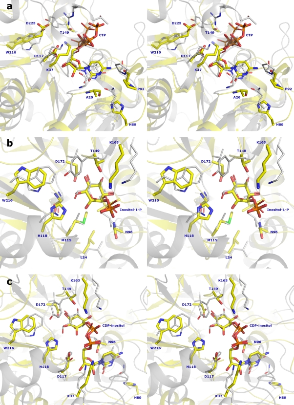
Fig. 5.
Structural superposition in stereo view of IPCT fitted with CTP and RmlA with dTTP (1G2V) (a), IPCT fitted with inositol-1-P and RmlA with G-1-P (1G23) (b), and IPCT structure fitted with CDP-inositol and RmlA with dTDP-d-glucose (1G1L) (c). The IPCT ribbon is drawn in yellow and RmlA in gray, with ligands shown as sticks (same atom color code as in Fig. 4, with sulfur atoms colored green and carbon atoms of RmlA colored gray).

