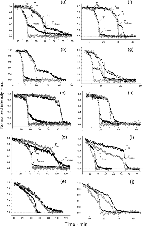
Fig. 2.
Germination of individual spores of various B. subtilis strains in response to l-alanine measured simultaneously by Raman spectroscopy and differential interference contrast (DIC) microscopy. (a to j) Spores of various strains of B. subtilis germinated at 25°C (a to e) or 45°C (f to j) in response to the addition of l-alanine. The dynamics of the germination of individual spores were monitored by both Raman spectroscopy (○) and DIC microscopy (●), and the times of various points in germination were determined as described in Materials and Methods. In each graph, time (in minutes) is shown on the x axis, and normalized intensity are shown in arbitrary units (a.u.) on the y axis. Note that data from only two individual spores are shown in panels a to j and that the two most adjacent curves with filled and open circles in all panels are from the same individual spore. The spores from strains PS533 (wild-type) (a and f), PS3411 (↑SpoVA) (b and g), FB62 (gerD) (c and h), PS3640 (spoVA1) (d and i), and PS3642 (spoVA2) (e and j) were analyzed and are shown in the various panels. In panels a and f, the times of Tlag, T1, and Trelease are indicated by arrows, but in panels d and i, only Tlag and Trelease are indicated, since T1 points could not be identified in most spoVA1 and spoVA2 spores germinating in response to l-alanine as described in Materials and Methods. The Trelease points in all panels are also noted by vertical dotted lines, and the horizontal dotted line at the bottom of the panels is the zero level as described in Materials and Methods.

