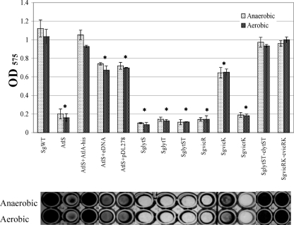
Fig. 8.
Biofilm formation assay. Cultures were grown in 1/4-strength BHI medium supplemented with 10 mM sucrose under aerobic or anaerobic conditions for 48 h. The top panel is quantification of the crystal violet-stained biofilms as detailed in the text. Data are representatives of the means of results of at least two separate experiments that were performed in triplicate, with error bars delineating standard deviations. *, statistically significant differences between the wild-type and mutant strains cultured under the same conditions (P < 0.05 [Student t test]). The bottom panel shows representative biofilms corresponding to the sample quantified in the bar graph above.

