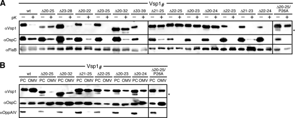
Fig. 4.
Localization of Vsp1 mutants. (A) Proteinase K (pK) accessibility immunoblots of Vsp1 tether mutants compared with that of OspCwt. FlaB was used as a periplasmic, protease-resistant control. (B) Membrane fractionation immunoblots of proteinase K-resistant, i.e., periplasmic Vsp1 tether mutants compared with that of OspCwt. OppAIV served as the IM control. OMV, outer membrane vesicle fraction; PC, protoplasmic cylinder fraction (also containing intact cells) (59, 64). An asterisk (*) in both panels indicates a CtpA-dependent OspC band (see text) (43) (Kumru et al., submitted).

