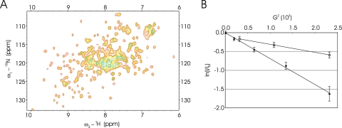
Fig. 3.
Examination of SsoHflX by NMR spectroscopy. (A) 2-D 1H-15N SOFAST-HMQC of SsoHflX at 50°C; (B) pulsed-field gradient diffusion NMR characterization of SsoHfIX (diamonds) compared to the 50S ribosomal subunit (open circles) at 25°C. The logarithm (y axis) of normalized resonance intensity (I/I0) is plotted against G2, the square of the gradient strength (x axis). I0 is the intensity at low gradient strength. The diffusion profile of SsoHflX is homogeneous, as indicated by the linear regression of ln(I/I0) as a function of G2. The diffusion coefficient extracted from the slope of ln(I/I0) as a function of G2 is 1.1 × 10⁻¹⁰ m2 s−1. The diffusion profile of the 50S resonances also appears to be homogeneous and corresponds to a diffusion coefficient of 4.2 × 10⁻¹¹ m2 s−1. Error bars indicate the uncertainty of the signal intensity based on the spectral noise.

