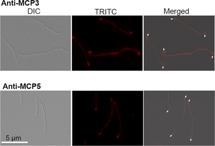
Fig. 2.
Localizations of MCP3 and MCP5 by using IFA. Wild-type cells were fixed with methanol, stained with either anti-MCP3 or anti-MCP5 antibody, and counterstained with anti-rat Texas Red antibody. The micrographs were taken under DIC light microcopy or fluorescence microscopy with a tetramethylrhodamine isothiocyanate (TRITC) emission filter (magnification, ×1,000), and the resulting images were then merged. Arrows point to the cellular locations of MCP3 and MCP5.

