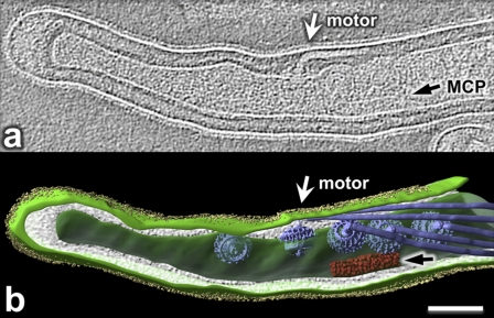
Fig. 4.
Cellular architecture of intact B. burgdorferi revealed by cryo-ET. (a) One central slice of a tomographic reconstruction from one organism displays the PF (white arrow) and MCP array (black arrow). (b) A 3-D model was generated by manually segmenting the outer membrane (light green), cytoplasmic membrane (green), flagellar filaments (blue), MCP array (red), and outer surface proteins (Osps) (yellow). 3-D maps of flagellar motors were computationally mapped back into the cytoplasmic membrane (26). Bar, 100 nm.

