Fig. 3.
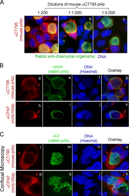
Detection of CT795 in the cytosol of C. trachomatis-infected cells. HeLa cells infected with C. trachomatis organisms were processed for immunolabeling with mouse antibodies visualized with a goat anti-mouse IgG conjugated with Cy3 (red), rabbit antibodies visualized with a Cy2-conjugated goat anti-rabbit IgG (green), and the DNA Hoechst stain (blue). (A) A mouse antibody raised against the GST-CT795 fusion protein was costained at various dilutions with a rabbit anti-chlamydia organism antibody. As the dilution of the anti-GST-CT795 antiserum increased, a unique cytosolic signal became clear (images b and c; red arrows). (B) The anti-GST-CT795 antibody (at 1:1,000) was costained with a rabbit anti-IncA antibody, and the CT795 signal (images d to g; red arrows) was detected outside the inclusion with a distribution pattern similar to that of CPAF (images h to k). (C) The costaining of the anti-CT795 antibody with the anti-chlamydial organism antibody was visualized under a confocal microscope. The CT795 signal (images l and o; red arrows) did not overlap with the organism signal (images m and o; green) at all. As a positive control, the CPAF-specific signal was also distinct from the organism signal in parallel cultures (images p to s).

