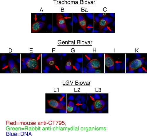
Fig. 7.
Secretion of CT795 into host cell cytosol is a common feature of all C. trachomatis serovars tested. HeLa cells infected with C. trachomatis trachoma biovar (serovars A, B, Ba, and C), genital biovar (D, E, F, G, H, I, and K), and LGV biovar (L1 to L3) as indicated on the top of each image were processed 40 h after infection for immunofluorescence labeling as described for Fig. 1. The CT795 antibody detected significant signals in the cytosol of host cells infected with all serovars from the Q3 biovars. Red arrows mark the CT795-sepcific signals in the cytosol of the chlamydia-infected cells.

