Fig. 9.
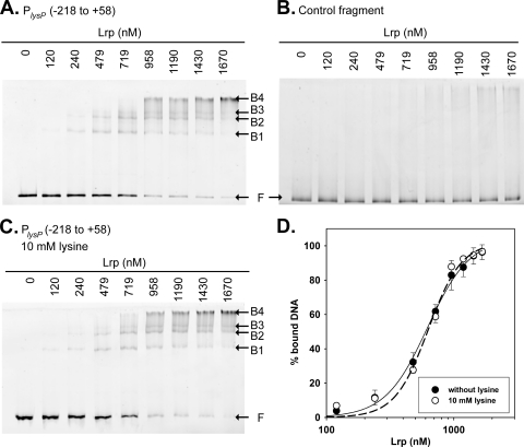
Lrp binding to the lysP promoter region. (A and C) Electrophoretic mobility shift assays (EMSAs) of the fluorescently labeled PlysP fragment (positions −218/+58) with increasing concentrations of purified His6-Lrp in the absence (A) or presence (C) of lysine. (B) EMSA performed with a DNA fragment within the lysP coding sequence as a control for unspecific binding. The positions of free DNA (F) and Lrp-DNA complexes (B1 to B4) are marked. (D) Binding profiles obtained after the quantification of free DNA and Lrp-bound DNA in EMSA gels were fitted using the Hill equation.

