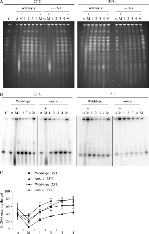
Fig. 3.
Branched DNA structures persist in rmi1-1 cells after a perturbed S phase. (A) Chromosome integrity is impaired following MMS treatment in rmi1-1 cells. Samples were taken from strains in Fig. 2C, and analyzed at the indicated times by pulsed-field gel electrophoresis (PFGE). C, yeast chromosomal DNA marker; α, α-factor-arrested sample; + M, sample taken after 1.5 h exposure to 0.0167% MMS; − M, sample taken after 50 min in the absence of MMS. 1, 2, 3 and 4 refer to the number of hours after the MMS was removed from the cultures. (B) Branched structures impair chromosome III electrophoretic mobility following MMS treatment in rmi1-1 cells. The DNA was transferred by Southern blotting and then hybridized with a probe that binds to ARS305 on chromosome III. (C) Quantification of chromosome III migration. The intensity of chromosome III in the wells versus the gel in panel B was quantified for each time point. The data are from three independent experiments. Error bars show standard errors.

