Fig. 5.
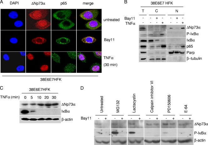
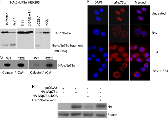
IKKβ affects stability and intracellular localization of ΔNp73α. (A) 38E6E7HFK cells were treated with TNF-α or Bay11 and ΔNp73, and p65 cellular localization was visualized by immunofluorescence with indicated antibodies. (B) 38E6E7HFK cells treated with TNF-α or Bay11 were collected and fractionated by using a nuclear extraction kit (Panomics). After fractionation, total extract (T), cytoplasm (C), and nucleus (N) were analyzed by immunoblotting with the indicated antibodies. (C) 38E6E7HFK cells were treated with TNF-α at the indicated time points. Protein extracts were prepared and analyzed by immunoblotting using the indicated antibodies. (D) 38E6E7HFK cells were treated with the indicated protease inhibitors for 8 h and Bay11 (+) or DMSO (−) for 2 h. Protein extracts were analyzed by immunoblotting. (E) HEK293 cells were transfected with the pcDNA3 HA-ΔNp73 construct and cultured under specified conditions (left and right panels) or cotransfected with different pcDNA3 constructs in the indicated combinations (right panel). Cells were then treated with indicated inhibitors followed by immunoprecipitation of whole-cell lysates and immunoblotting with the indicated antibodies. (F) 38E6E7HFK cells were seeded on coverslips and treated for 8 h with E-64 and/or for 2 h with Bay11. After the cells were fixed, ΔNp73 was visualized by immunofluorescence with specified antibodies. (G) In vitro-translated HA-tagged WT and mutant 422E ΔNp73 were incubated with recombinant calpain I in the presence or absence of Ca2+. After 30 min, the reaction was stopped by the addition of SDS loading buffer, and samples were analyzed by immunoblotting with an anti-HA-tag antibody. (H) 38E6E7HFK cells were transfected with the pcDNA3-expressing wild type or 422A or 422E HA-ΔNp73α. After 24 h, protein extracts were analyzed by immunoblotting with the indicated antibodies.


