Fig. 7.
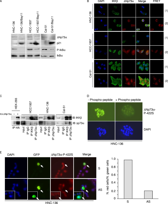
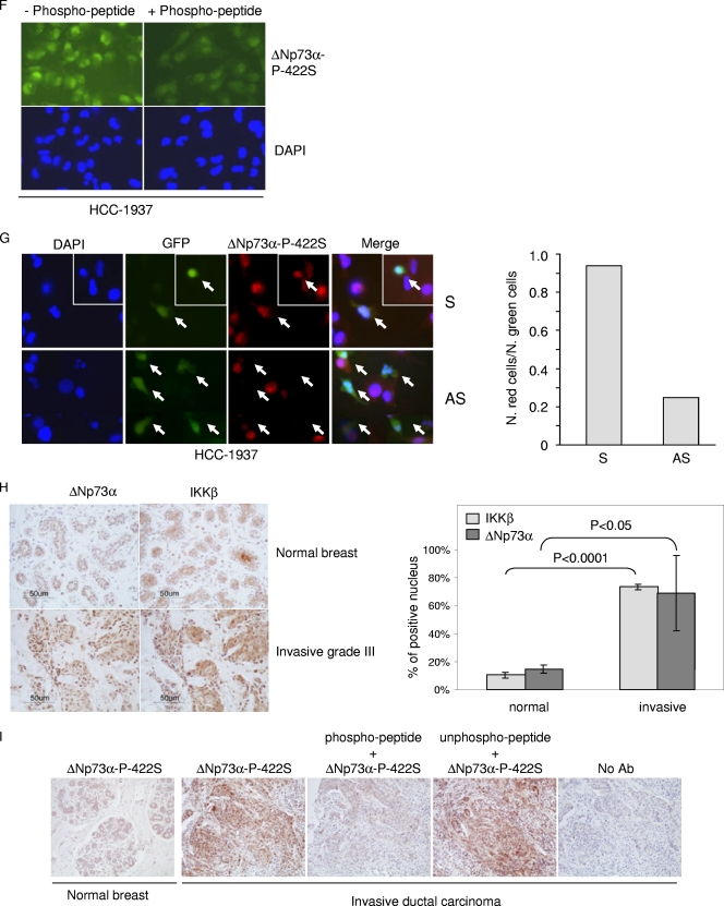
IKKβ and ΔNp73α cross talk in cancer-derived cell lines and primary cancers. (A) HNC-136, HCC-1937, and Cal-51 cell lines were treated with Bay11 for 2 h, and total protein extracts were analyzed by immunoblotting with the indicated antibodies. (B) HCC-1937, HNC-136, and Cal-51 cells were stained with the indicated antibodies. Fluorescent staining was visualized by confocal microscopy. FRET positive signals (2) and FRET negative controls (1) are shown in the right panels. (C) One-milligram protein extracts of HCC-1937, HNC-136, and Cal-51 were immunoprecipitated with an anti-IKKβ or -ΔNp73α antibody. As negative controls, the same amounts of total extracts from each cell line were immunoprecipitated with anti-IgG. Immunocomplexes were analyzed by immunoblotting. One-tenth of total extracts used for the immunoprecipitation was loaded as input. As a control, immunocomplexes obtained from HEK293 cells transfected with pcDNA or pcDNA3-HA-ΔNp73α were included in the experiment. (D) HNC-136 cells were stained with the indicated antibodies. Fluorescent staining was visualized by confocal microscopy. As a control, primary antibody was preincubated with an excess of 422S-phosphorylated peptide. (E) HNC-136 cells were cotransfected with pE-GFPCI and S or AS against ΔNp73α. Thirty hours posttransfection, cells were stained with anti-ΔNp73α P-422S antibody and analyzed for immunofluorescence. Representative images are shown in the left panel; arrows indicate transfected cells. The white frame indicates a different field. Quantification of ΔNp73α P-422S and GFP-positive cells or GFP-positive cells was determined by counting at least 100 transfected cells in more than 10 different fields (right panel). (F) HCC-1937 cells were stained as explained in the legend for panel D. (G) HCC-1937 cells were transfected and processed as explained in the legend for panel E. Representative images are shown in the left panel; arrows indicate transfected cells. The white frame indicates a different field. Quantification of ΔNp73α P-422S and GFP-positive cells or GFP-positive cells was determined by counting at least 100 transfected cells in more than 10 different fields (right panel). (H) ΔNp73 and IKKβ cellular localization was analyzed in normal and cancer breast tissues (left panel, representative staining). The histogram (right panel) shows the quantification of the percentage of cells in normal (n = 1) or cancer tissues (n = 3) with nuclear staining for IKKβ and ΔNp73α. (I) Normal and breast cancer tissues were stained with a specific antibody against the 422S-phosphorylated form of ΔNp73α. As controls, immunostaining was also performed without the primary antibody (no Ab) or with primary antibody preincubated with a phosphorylated or nonphosphorylated 422S ΔNp73α peptide.


