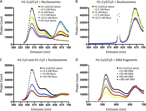
Fig. 4.
FRET analysis of H1 binding to nucleosomes indicates intramolecular folding of the CTD. (A) Emission spectra of H1 (10 nM) labeled at either end of the CTD as described in Materials and Methods in the absence of nucleosomes (black circles) or in the presence of 2.5, 5.0, 7.5, or 12.5 nM 217N nucleosomes, as indicated. The spectrum of H1 labeled exclusively with Cy3 is also shown (brown line). Excitation was at 515 nm. (B) As in panel A except with excitation at 610 nm. (C) Nucleosome-induced FRET is entirely intramolecular. H1 was labeled exclusively with either Cy3 or Cy5 and then mixed in a 1:1 ratio, and emission spectra were examined in the absence of nucleosomes (black circles) or in the presence of 2.5, 5, or 10 nM nucleosomes. (D) FRET of H1 labeled with both Cy3 and Cy5 (as in panel A) was determined in the presence of increasing amounts of 207-bp naked 601 DNA fragments with excitation at 515 nm. Corresponding emission spectra for excitation at 610 nm are not shown.

