Fig. 6.
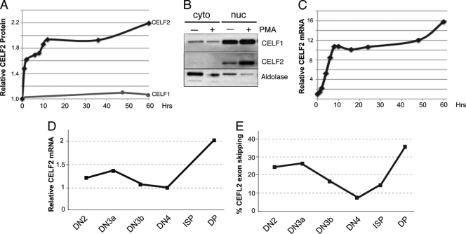
Expression of CELF2 in JSL1 cells and thymocytes. (A) Quantification of total CELF2 and CELF1 protein in JSL1 cells following stimulation with PMA. Protein level was determined by Western blotting and normalized to actin. (B) Representative Western blot of CELF2 and CELF1 as used for panel A at 48 h following stimulation. Cells were fractionated upon lysis and blotted for adolase as a measure of the purity of cytoplasmic (cyto) and nuclear (nuc) fractions. Results indicate that while expression of CELF2 changes upon stimulation, subcellular localization of this protein is not altered. (C) Quantification of total CELF2 mRNA in JSL1 cells following stimulation with PMA. mRNA was determined by RT-PCR and normalized to glyceraldehyde-3-phosphate dehydrogenase (GAPDH). (D) Quantification of CELF2 mRNA in murine thymic populations. Analysis was done as described above for panel C. (E) Quantification of percent exon skipping in CELF2 mRNA in the indicated murine thymic populations, showing a sharp rise in skipping between DN4 and DP, where the increase in mRNA and presumably protein is also observed.

