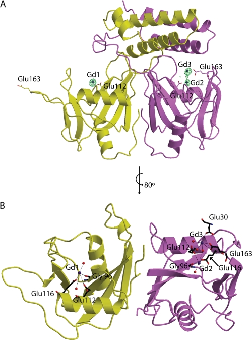
Fig. 5.
Gd3+-bound CDP-Chase structure. In both panels, the coloring of the protein monomers is similar to that in Fig. 2. (A) The structure of the enzyme with Gd3+ (blue spheres) is shown with an anomalous difference map contoured at 5σ (green mesh). Residues mutated are drawn in sticks. (B) The coordination of these Gd3+ ions is shown. (For simplicity, the N-terminal domain was omitted in this panel).

