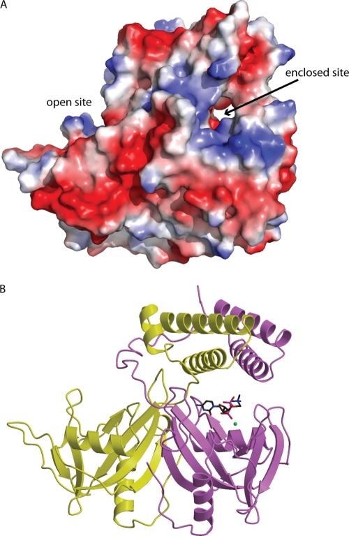
Fig. 7.
Model of CDP-choline binding. (A) An electrostatic surface representation of B. cereus CDP-Chase showing the narrow channel comprising the enclosed site and the open cavity comprising the open site. (B) The energy-minimized complex of CDP-Chase and CDP-choline (sticks). The modeled Mg2+ is shown as a green sphere.

