Fig. 1.
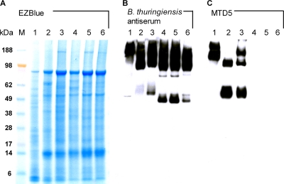
Immunodetection of anthrose-containing tetrasaccharide in B. anthracis strains isolated from cattle carcasses in African countries. (A) EZBlue (Sigma) protein staining of B. anthracis spore lysates (lane 1, Sterne strain; lane 2, internal reference strain JF3783 from Switzerland; lane 3, isolate from Zimbabwe; lane 4, isolate from Mali; lane 5, one representative isolate from Chad; lane 6, one representative isolate from Cameroon) separated on a 4 to 10% gradient SDS-polyacrylamide gel. (B) Western blot analysis of the reactivity of anti-B. thuringiensis antiserum diluted 1:1,000 (27). (C) Cross-reactivity of the anti-anthrose-rhamnose disaccharide MAb MTD5 with the blotted total spore lysates. Similar results were obtained with all other antidisaccharide (MTD1 to MTD4 and MTD6) and antitetrasaccharide (MTA1 to MTA3) MAbs (14, 26–28) tested. MAb MTD5 was used at a concentration of 0.01 μg/ml. The sizes of the molecular mass markers (lane M) are given in kDa.

