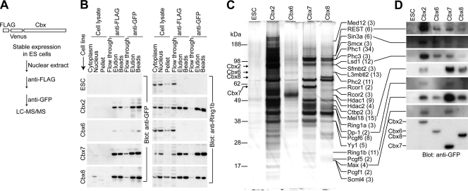
Fig. 1.
Purification of proteins associated with Cbx family members from mouse ES cells. (A) Flow diagram for immunoaffinity purification and identification of proteins associated with Cbx family members from mouse ES cells that expressed Cbx fusion proteins (43). (B) The enrichments of Cbx fusion proteins and of Ring1b during purification from the cell lines indicated to the left of each blot were evaluated. An equal proportion of each fraction (indicated above the lanes) was analyzed by immunoblotting using anti-GFP (left) and anti-Ring1b (right) antibodies. Multiple exposures of the blots were scanned, and the total protein concentrations were measured to calculate the fold enrichment of each Cbx protein and of Ring1b at each stage of purification. The final fold enrichments of each Cbx protein and of Ring1b were 310,000 and 240 (Cbx2), 1,000 and 1.5 (Cbx6), 12,000 and 1,000 (Cbx7), and 6,600 and 390 (Cbx8), respectively, relative to the cell lysates. ESC, parental ES cell line used to generate the ES cell lines expressing Cbx fusions. (C) SDS-PAGE analysis of the fractions purified from cells that expressed the Cbx fusion proteins indicated above the lanes or the parental cells (ESC). The mobilities of PRC1- and REST-associated proteins identified by mass spectrometry are indicated by the labels at the right of the images. The number of unique peptides corresponding to each protein is indicated in parentheses after the name of each protein. (D) Fractions purified from cells that expressed the Cbx fusions indicated above the lanes were analyzed by immunoblotting using antibodies directed against the proteins connected to the lines on the left of the images. The Cbx fusions were detected by using anti-GFP antibodies (bottom). Equal proportions of the fractions purified from cells that expressed Cbx2, Cbx7, and Cbx8 fusions and from the parental cells (ESC) were analyzed. A 10-fold-larger proportion of the fraction purified from cells that expressed the Cbx6 fusion was analyzed to compensate for the loss of the Cbx6 fusion during the isolation of nuclei.

