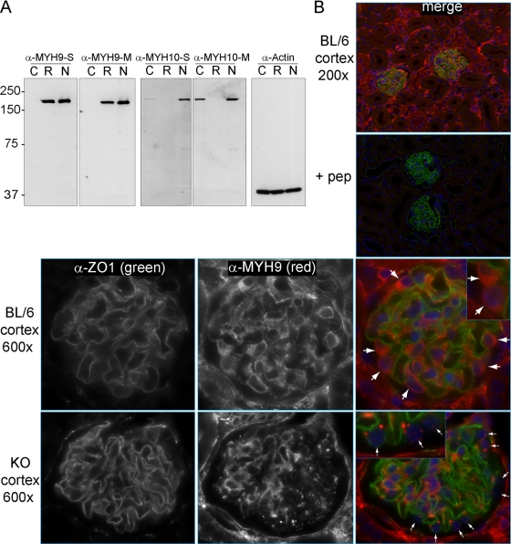
Fig. 2.
Localization of MYH9 in mouse kidneys. (A) Immunoblots to characterize new anti-MYH9 and anti-MYH10 antibodies, comparing equivalent titers of crude serum (S) or Melon gel-purified antibody (M). Lysates: C, COS7 cells (which do not express MYH9); R, rat basophil leukemia cells (which do not express MYH10); and N, NIH 3T3 cells (which express both MYH9 and MYH10). (B) Indirect IF with purified anti-MYH9 (1:400) and anti-ZO1 (1:200) in perfusion-fixed kidneys. (Top right) The panel (merge) is a ×200 view demonstrating ZO1 in glomeruli and MYH9 in a subset of tubules as well as glomeruli. (Top right, second panel) MYH9 peptide (1 ng/μl) was added to the anti-MYH9 and anti-ZO1 primary Ab mix (+ pep), and with identical photo settings, there was no MYH9 staining, while ZO1 staining was unchanged. Secondary Ab-only controls were similarly dark. (Top row) A ×600 view of a glomerulus demonstrates anti-MYH9 staining of podocyte cytoplasm (large white arrows) as well as mesangial and possibly endothelial cells within the glomerulus. (Bottom row) In glomeruli from KO mice, MYH9 is no longer visible in podocytes (small white arrows), while MYH9 staining of the mesangium and other cells is unchanged.

