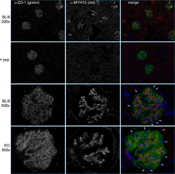
Fig. 3.
Localization of MYH10 in mouse kidneys. Indirect IF was performed with anti-MYH10 (1:800), as characterized by immunoblotting in Fig. 2A, and anti-ZO1 (1:200). (Top row) Images at a magnification of ×200 show MYH10 in glomeruli and rare tubules. (Second row) MYH10 peptide (0.2 ng/μl) was added to the anti-MYH10 and anti-ZO1 primary Ab mix (+ pep). With identical photo settings, MYH10 staining was competed away while ZO1 staining was unchanged. (Third row) Images of glomeruli at a magnification of ×600 reveal MYH10 in mesangial cells but not in podocytes (small white arrows). (Bottom row) MYH10 expression in kidneys of KO mice is unchanged from that in controls, with prominent mesangial staining and no staining of podocytes (small white arrows).

