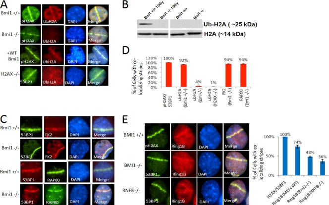
Fig. 5.
(A) Bmi1+/+; Ink4a−/−; Arf−/− MEFs, Bmi1−/−; Ink4a−/− Arf−/− MEFs, Bmi1−/−; Ink4a−/− Arf−/− MEFs reconstituted with wt-BMI1, or H2AX−/− MEFs were treated with laser scissors and, after 30 min, processed for IF using antibody to Ub-H2A-K119 and either 53BP1 or pH2AX as indicated. (B) Bmi1+/+; Ink4a−/−; Arf−/− MEFs or Bmi1−/−; Ink4a−/− Arf−/− MEFs were treated with 10 Gy of IR or mock treated and, after 30 min, chromatin extracts were prepared and processed for Western blotting with antibody to Ub-H2AK119 or antibody to histone H2A. (C) Bmi1−/−; Ink4A−/− Arf−/− MEFs were treated with laser scissors and, after 30 min, processed for IF with antibodies to polyubiquitin (FK2) or RAP80 and 53BP1 as indicated. (D) Bar graphs show the percentages of the indicated cell types with laser scissors-induced regions of pH2AX or 53BP1 that also show colocalization with Ub-H2A-K119, FK1, or RAP80. (E) Bmi1+/+; Ink4a−/− Arf−/− MEFs, Bmi1−/−; Ink4a−/− Arf−/− MEFs, or Rnf8−/− MEFs were treated with laser scissors and, after 30 min, processed for IF using antibodies to RING1B and either pH2AX or 53BP1 as indicated. The percentages of cells with laser scissors-induced damage showing colocalization of RING1B with pH2AX or 53BP1 for each MEF strain is shown in the bar graph. All data are plotted as means ± the SEM of at least three independent experiments.

