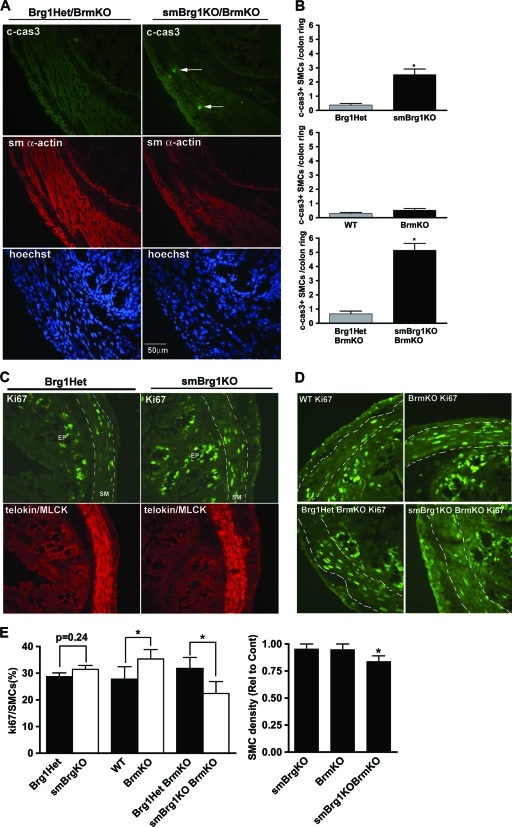
Fig. 8.
Alterations in apoptosis and proliferation in colon smooth muscle from knockout mice. (A) Cleaved caspase 3 (green), SM α-actin (red), and Hoechst nuclear (blue) staining of colons from neonatal smBrg1/Brm double knockout and control Brg1Het/Brm knockout mice. Arrows point to cleaved caspase 3-positive SMCs. (B) Cleaved caspase 3-positive SMCs in the circular muscle layer of colons from knockout mice were counted and quantitated. Significant differences between knockout and control mice were determined by Student's t test. *, P ≤ 0.05. Data are the means ± SEM from 3 to 5 mice. (C and D) Ki67 (green) and telokin/MLCK (red) immunofluorescent staining of colon from neonatal smBrg1 knockout (C) and Brm knockout and smBrg1/Brm double knockout (D) mice. (E) Left panel, Ki67-positive SMCs were quantitated in stained sections similar to those shown in panels C and D except that nuclei were also stained with Hoechst stain. Right panel, smooth muscle cell density in the circular muscle layer of colons from knockout mice expressed relative to the appropriate control mice (indicated in panels C and D). Data are the means ± SEM from 5 or 6 mice. *, P ≤ 0.05.

