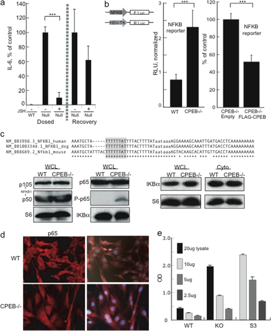
Fig. 4.
NF-κB p65 is constitutively active in CPEB KO MEFs. (a) WT and CPEB KO MEFs were incubated with the NF-κB inhibitor JSH-23 or the vehicle alone for 48 h. After 48 h, the cells were washed and cultured in fresh medium without JSH-23. Samples of the medium were taken at 48 h, and IL-6 was determined by ELISA. (b) MEFs were transfected with a firefly NF-κB-promoter reporter and a Renilla luciferase reporter for normalization into WT, CPEB KO, or CPEB KO cells transduced with FLAG-CPEB or the empty vector. Five days later, the luciferase expression was determined. (c) Alignment of a portion of the 3′ UTR of human, dog, and mouse NF-κB1. The putative CPE is highlighted, and the polyadenylation hexanucleotide is in lowercase. WCL, whole-cell lysate; Cyto, cytoplasm. The Western blots show the relative levels of various NF-κB family members, as well as the regulatory protein IKBα. P-p65 refers to p65 phosphorylated on residue 276. S6 served as a loading control. (d) Localization of p65 in WT and CPEB KO MEFs determined by immunocytochemistry analysis. The cells were also stained with DAPI and merged. (e) NF-κB p65 transcription factor binding assay. The indicated amounts of nuclear lysate from WT, CPEB KO, and HeLa S3 cells were mixed with a biotinylated oligonucleotide containing the NF-κB consensus binding site. Antibody against p65 was then used to determine the amount of this protein captured by streptavidin. Asterisks indicate P values of <0.001. OD, optical density.

