Fig. 5.
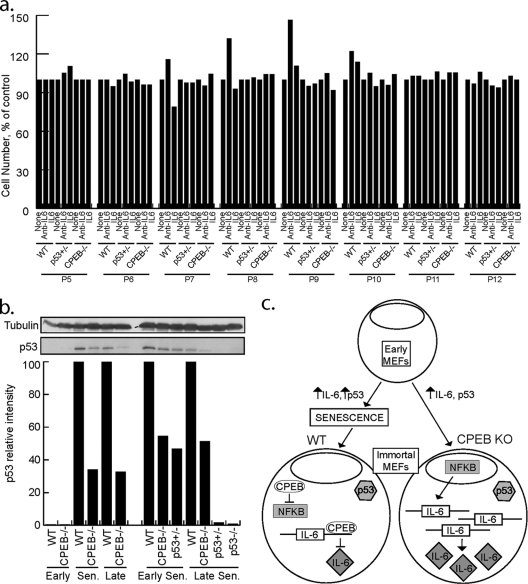
A senescence regulatory pathway composed of IL-6, CPEB, and p53. (a) WT, p53 heterozygous, or CPEB KO MEFs were incubated in the presence of IL-6 neutralizing antibody or recombinant IL-6. The agents were added to the culture medium and replenished at each passage, and this was followed by determination of cell numbers. (b) p53 protein levels are reduced in CPEB KO MEFs. Lysates prepared from WT, CPEB KO, or p53 heterozygous MEFs at various passage points were analyzed by Western blotting for p53 and quantified. β-Tubulin served as a loading control. Sen, senescence. (c) Model illustrating interplay among CPEB, IL-6, NF-κB, and p53 in controlling senescence. Early WT and CPEB-null MEFs exhibit low levels of p53 and IL-6. As WT and CPEB KO MEFs increase in passage number, IL-6 levels increase. In WT MEFs, higher levels of IL-6 reinforced the senescence response, p53 protein levels increase, and cells enter senescence and have reduced division rates. In contrast, CPEB KO MEFs do not enter senescence and do not exhibit decreased division and p53 levels are 50% of those of passage-matched WT MEFs, likely through lack of CPEB-mediated polyadenylation of p53 mRNA. After WT cells become immortalized, IL-6 levels are decreased and NF-κB is not active. In contrast, passage-matched immortalized CPEB KO MEFs continue to secrete high levels of IL-6 and NF-κB is activated. In immortalized MEFs, p53 is likely mutated. Immortalized CPEB KO MEFs continue to secrete IL-6 for many passages.

