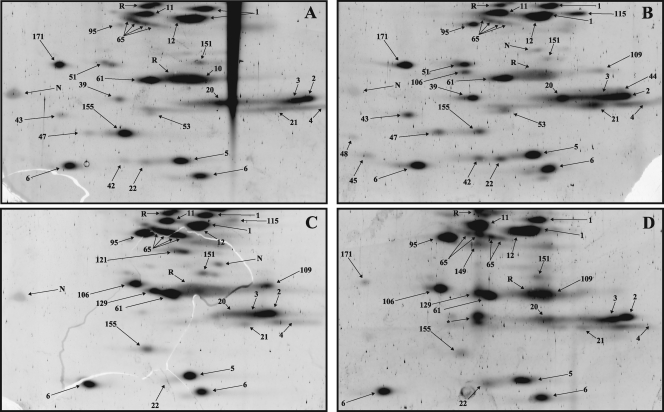
Fig. 8.
2D gel electrophoresis of low-molecular-mass proteins from affinity-purified spliceosomal A (A), B (B), Bact (C), and C (D) complexes. Spliceosomal complexes formed on PM5-based pre-mRNA were analyzed by 2D gel electrophoresis using conditions optimal for the separation of proteins under 25 kDa in mass, and spots were visualized by staining with Sypro Ruby. None of the low-molecular-mass proteins were detected at levels significantly above background upon staining with Pro-Q Diamond (data not shown). Spot no. 171 corresponds to ALG-2/PDCD6 (gi∣121948367) and was abundant only in B complexes formed on the PM5 pre-mRNA.

