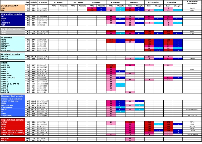
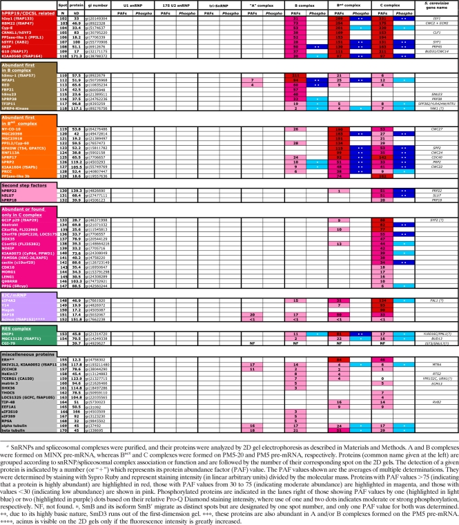
Table 1.
Abundances and phosphorylation statuses of proteins detected in the major spliceosomal snRNPs and spliceosomal A, B, Bact, and C complexesa
SnRNPs and spliceosomal complexes were purified, and their proteins were analyzed by 2D gel electrophoresis as described in Materials and Methods. A and B complexes were formed on MINX pre-mRNA, whereas Bact and C complexes were formed on PM5-20 and PM5 pre-mRNA, respectively. Proteins (common name given at the left) are grouped according to snRNP/spliceosomal complex association or function and are followed by the number of their corresponding spot on the 2D gels. The detection of a given protein is indicated by a number (or “+”) which represents its protein abundance factor (PAF) value. The PAF values shown are the averages of multiple determinations. They were determined by staining with Sypro Ruby and represent staining intensity (in linear arbitrary units) divided by the molecular mass. Proteins with PAF values >75 (indicating that a protein is highly abundant) are highlighted in red, those with PAF values from 30 to 75 (indicating moderate abundance) are highlighted in magenta, and those with values <30 (indicating low abundance) are shown in pink. Phosphorylated proteins are indicated in the lanes right of those showing PAF values by one (highlighted in light blue) or two (highlighted in purple) dots based on their relative Pro-Q Diamond staining intensity, where use of one and two dots indicates moderate or strong phosphorylation, respectively. NF, not found. *, SmB and its isoform SmB′ migrate as distinct spots but are designated by one spot number, and only one PAF value for both was determined. **, due to its highly basic nature, SmD3 runs out of the first-dimension gel. ***, these proteins are also abundant in A and/or B complexes formed on the PM5 pre-mRNA. ****, acinus is visible on the 2D gels only if the fluorescence intensity is greatly increased.


