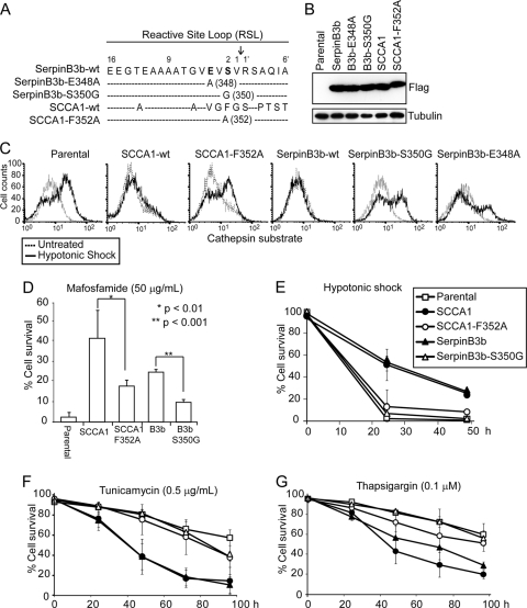
Fig. 3.
SCCA1's ability to enhance ER stress-induced cell death requires its protease-inhibitory activity. (A) Diagram of the location of point mutations made within the reactive site loop of SCCA1 and SerpinB3b. The numbering indicates the position of residues within the reactive site loop, and the arrow indicates the protease cleavage site. (B) Immunoblot of bax−/− bak−/− BMK cells stably expressing Flag-tagged wild-type (wt) and mutant SCCA1 and SerpinB3b. (C) Indicated cells were treated with hypotonic shock for 6 h and then incubated with a fluorogenic cathepsin L substrate. Cathepsin L activity was determined by flow cytometry. (D to G) Parental cells or cells expressing the indicated proteins were treated with mafosfamide for 36 h (D), hypotonic shock (E), tunicamycin (F), or thapsigargin (G) for the indicated amount of time. Cell death was measured by PI exclusion.

