Fig. 5.
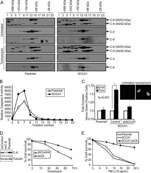
SCCA1 enhances caspase-8 oligomerization and activation in response to ER stress. (A) bax−/− bak−/− BMK parental and SCCA1-expressing cells were treated with tunicamycin (0.5 μg/ml) for 16 h, and size exclusion chromatography was performed. Eluted fractions were probed for caspases-8, -2, and -9. Molecular mass of the fractions is indicated at the top. (B) Caspase-8 activity in tunicamycin-treated fractions was determined using a luminogenic caspase-8 substrate, IETD. (C) Parental and SCCA1 cells expressing either an shControl or shSCCA1 were transfected with Venus-tagged caspase-8. Cells were left untreated or treated for 16 h with tunicamycin and then analyzed by flow cytometry. Shown is the fold increase in fluorescent cells in tunicamycin-treated cells compared to untreated cells. Inserted images show the Venus fluorescence induced by tunicamycin treatment. (D) Caspase-8 knockdown protects SCCA1-expressing cells from ER stress-induced apoptosis. SCCA1-expressing bax−/− bak−/− cells were infected with shCaspase-8 (shC-8) or control shRNA. The cells were treated with tunicamycin, and cell death was measured by PI exclusion. (E) Parental and SCCA1-expressing cells as well as SCCA1-expressing cells containing shCaspase-8 were treated with TNF-α plus cycloheximide (10 μg/ml), and cell death was measured by PI exclusion.

