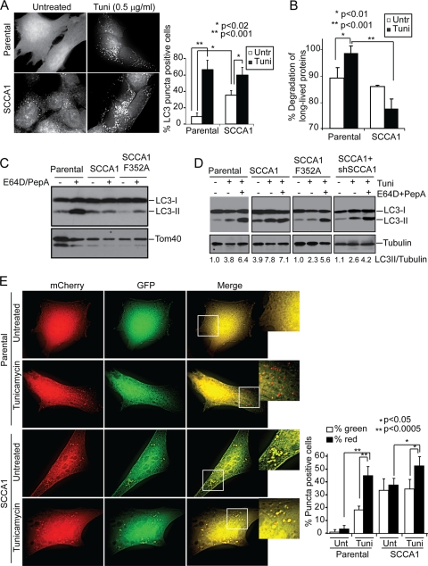
Fig. 7.
SCCA1 blocks lysosomal turnover. (A) bax−/− bak−/− BMK parental or SCCA1-expressing cells stably expressing GFP-LC3 were treated with tunicamycin (Tuni) for 8 h. Cells were observed under a deconvolution microscope. The percentage of cells showing punctated GFP-LC3 was determined for three independent counts of 50 cells, and the values represent the mean ± standard error of the mean. (B) Cells were labeled with [14C]valine for 48 h and then treated for 24 h with tunicamycin. Long-lived protein degradation was measured. Untr, untreated. (C) bax−/− bak−/− BMK parental cells or SCCA1- or SCCA1-F352A-expressing cells were left untreated or treated with E64D (10 μg/ml) and PepA (10 μg/ml) for 16 h. Cell lysates were probed for LC3. Tom40 was probed for equal loading. (D) Indicated cells were treated with tunicamycin alone or in combination with E64D (10 μg/ml) and PepA (10 μg/ml) for 16 h. Cell lysates were probed for LC3 and tubulin, and the ratio of LC3-II against tubulin was determined. (E) Parental and SCCA1-expressing bax−/− bak−/− BMK cells were transfected with mCherry-EGFP-LC3 and then treated with tunicamycin (0.5 μg/ml) for 16 h. The amount of red and green puncta was quantitated. Percentage of cells showing green and red puncta was determined for three independent counts of 50 cells, and the values represent the mean ± standard error of the mean.

