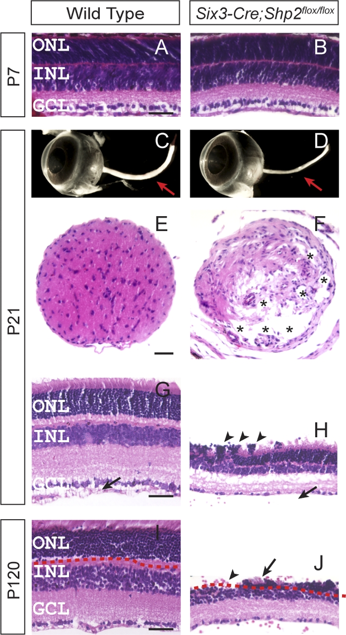
Fig. 1.

Severe degeneration of the retina and optic nerve in Six3-Cre; Shp2flox/flox adult mice. (A and B) Normal retinal histology in the Six3-Cre; Shp2flox/flox mutant at postnatal day 7 (P7). (C to F) In the Six3-Cre; Shp2flox/flox adult mice, the loss of optic nerve was observed in the dissected eyeballs (arrow in panel D) and in the transverse sections (asterisks indicate the vacuoles in the mutant optic nerve in panel F). (G to J) The Six3-Cre; Shp2flox/flox mutant retina sections were approximately 50% thinner than that of the wild type at P21 (H), and the outer nuclear layer (ONL) was completely absent in some retinal regions at P120 (the arrow in panel J indicates the neighboring residual photoreceptor cells). Notice the photoreceptor cell rosette (arrowheads in panel H) and the lack of the optic nerve fiber layer/inner limiting membrane layer (NFL/ILML) in the mutant retina (arrow in panel H). GCL, ganglion cell layer. Bars, 40 μm.
