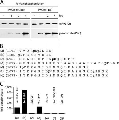
Fig. 6.
Identification of eIF4G phosphorylation sites by PKCα in vitro. (A) Time course of phosphorylation of recombinant GST-Ct by purified, recombinant GST-PKCα. This experiment was repeated four times; a representative assay is shown. (B) Sequences of phosphorylated eIF4G peptides determined from tandem mass spectrometry. Bold pS or pT denotes a phosphorylated Ser or Thr residue, respectively. (C) The relative intensities of phosphorylation of corresponding residues in eIF4G were determined by quantitative phospho-proteomic analysis of in vitro phosphorylated, recombinant GST-Ct by purified, recombinant GST-PKCα.

