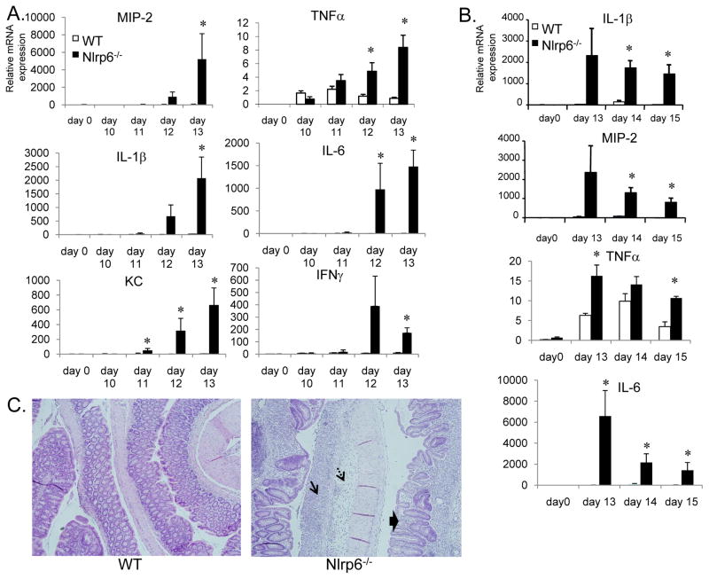
Figure 5. Increased proinflammatory mediator production and delayed resolution of inflammation in Nlrp6-deficient mice.
A, mRNA was isolated from age- and sex-matched wildtype and Nlrp6-deficient on days 0 (N=3 mice/group), 10–13 (N=5 mice/group/timepoint) after treatment with AOM/DSS. mRNA expression of various proinflammatory mediators relative to β-actin was measured by quantitative real-time PCR. B, mRNA expression of proinflammatory mediators on days 13 to 15 after the first round of DSS in the AOM/DSS tumor induction protocol. Data expressed as means +/− S.E.M., *, p< 0.05 by Student’s t-test. C, Representative photographs of hematoxylin and eosin-stained colon sections from Nlrp6−/− and wildtype (WT) mice on day 15. Single arrow shows area of mucosal ulceration; dashed arrows point to submucosa edema. Thick arrows show regenerating crypts in a background of inflammation with inflammatory cell infiltration.

