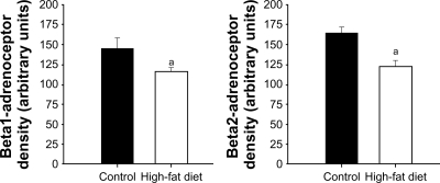
Figure 4.
Western blot analysis of β1- and β2-ARs obtained from control and high-fat-fed Ossabaw swine left ventricles. Western blot analyses revealed a significant decrease in ventricular β1- and β2-ARs: 19.86% and 22.6%, respectively. Signal intensities were normalized to concomitant β-actin.
Note: aP < 0.05 versus control.
Abbreviation: AR, adrenoceptor.

