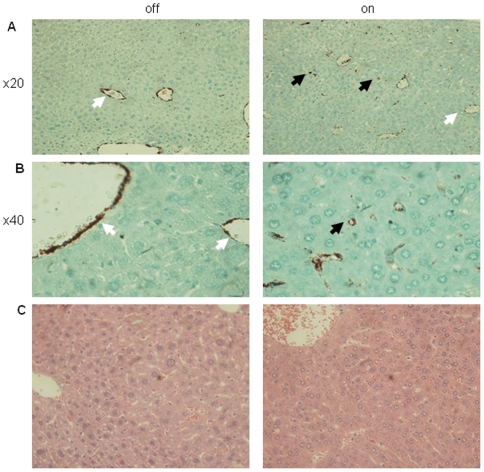
Figure 4. Sinusoidal capillarization in sVEGF-R1 expressing livers without parenchymal damage.
(a,b) Immunohistochemical staining for vWF (Von-Willebrand Factor) on liver sections (black arrows-sinusoids, white arrows-larger blood vessels). (c) H&E staining of liver sections showing normal appearance.

