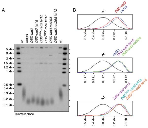
Figure 2.
Telomeric repeat length analysis of DBD-rad3 strains. (A) Southern blot hybridization analysis of ApaI digested genomic DNA, probed with telomeric repeat sequences. All strains were extensively streaked on agar plates prior to preparation of genomic DNA to ensure terminal telomere phenotype, and all lanes contained comparable genomic DNA based on ethidium bromide (EtBr) staining (data not shown). (B) Quantification of hybridization signal shown in (A) by ImageQuant software (Molecular Dynamics). Peaks of telomeric repeat hybridization signal were normalized and plotted against DNA size.

