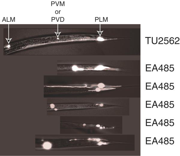
Figure 3.
Fluorescence micrographs showing PLM axonal morphology in TU2562 and EA485. The top panel shows a PLM with normal morphology in a TU2562 animal. The next five panels show examples of PLMs with axonal morphology defects in EA485 animals. The white spots anterior to the PLMs are the mec-3 expressing PVM and PVD neurons.

