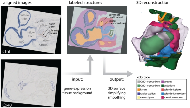
Figure 2. 3D reconstruction from sections.
The left panel shows two exemplary ISH-stains of expression of cTnI and Cx40, translated and rotated to alignment. Note that the line that indicates the splanchnic plexus and the textual annotations were later added for the sake of clarity. Using Amira, the signals of gene-expression and the general tissue background were transformed into a 3D reconstruction. The presented colors indicate the same structures throughout the remainder of the article. Note that yellow is used to indicate both the cardiac cushions after invasion with mesenchyme, but also for mesoderm that is of unclear splanchnic or somatic descent. (Abbreviations–3D: three dimensional; AVC: atrioventricular canal; cTnI: cardiac Troponin I; Cx40: Connexin 40.)

