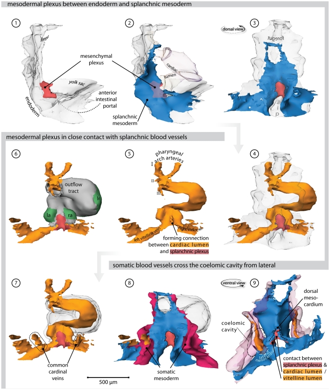
Figure 3. Heart region of stage 12 embryo.
Also refer to supplemental File S1 for an interactive version of this reconstruction. (1) Right lateral view of the endoderm (transparent). Overlying the endoderm of the anterior intestinal portal is a mesenchymal plexus (red). (2) Overlying this mesenchymal plexus is the splanchnic mesoderm (blue). The lumen of the heart tube is transparent. (3) Dorsal view, showing how the vascular plexus is wedged between the endoderm and the splanchnic mesoderm. (4) The cardiovascular lumen is shown in orange; the forming vitelline veins are continuous with the vessels that cover the yolk sac. (5) Cardiac lumen protrudes into dorsal mesocardium, where contact with splanchnic plexus is being established. (6) Bilaterally flanking the dorsal mesocardium are the emerging atria, as distinguished by expression of Connexin40 (green). (7) Hooking into the vitelline veins from dorsolateral are the common cardinal veins. (8) These common cardinal veins reside in somatic mesoderm (red). (9) Ventral view of the splanchnic and somatic mesoderm. Separating these tissues is the coelomic cavity. They contact at the so-called lateral mesocardium (light blue). (Abbreviations–I, II, III: first, second & third pharyngeal arch artery, respectively; aip: anterior intestinal portal; la: left atrium; ra: right atrium; lv: left ventricle.)

