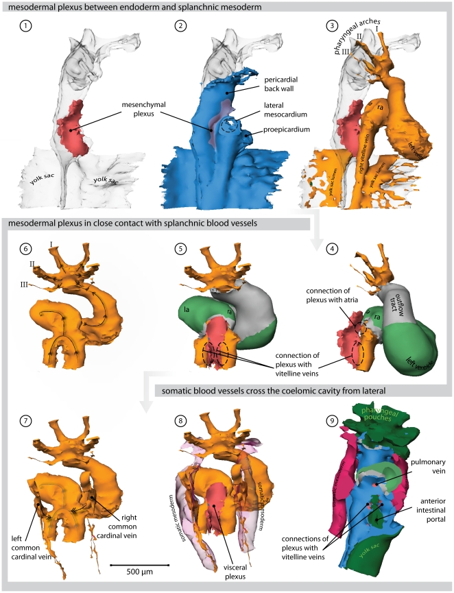
Figure 4. Heart region of stage 16 embryo.
Also refer to supplemental File S1 for an interactive version of this reconstruction. (1) Right lateral view of the endoderm (transparent). Overlying the endoderm of the anterior intestinal portal is a mesenchymal plexus (red). (2) Overlying this plexus is the splanchnic mesoderm (blue); its connection with the somatic mesoderm, the lateral mesocardium, is removed. (3) Cardiovascular lumen in relation with the endoderm and splanchnic plexus. (4) The lumina of the blood islands covering the yolk sac are removed. Primary myocardium is shown in grey, Cx40 positive working myocardium in green. The splanchnic plexus is separating and contacts both the atrial lumen via the dorsal mesocardium, and the vitelline veins. (5) Dorsal view of the plexus in relation to the myocardium and the vitelline veins. (6) The direction of blood-flow within the heart and vitelline vessels is indicated with arrows. (7) Addition of the cardinal veins. (8) The cardinal veins reside in somatic mesoderm (transparent red). (9) Ventral view, now showing the endoderm in dark green. The surface of the reconstructions is cut in a frontal plane, showing canalization of the splanchnic plexus into the cardiovascular lumen of the atria (via the dorsal mesocardium) and the vitelline veins. (Abbreviations–I, II, III: first, second & third pharyngeal arch artery, respectively; la: left atrium; ra: right atrium.)

