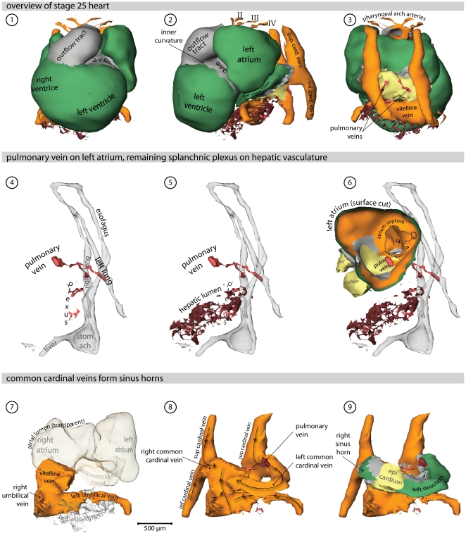
Figure 6. Heart region of stage 25 embryo.
Also refer to supplemental File S1 for an interactive version of this reconstruction. (1) Ventral view of the heart. Cx40 positive working myocardium is shown in green, primary myocardium is depicted in grey. (2) Left lateral view of the heart; the splanchnic plexus is shown in red and hepatic lumen in brown. (3) Dorsal view of the heart. (4) Left lateral view of the endoderm (transparent grey) in relation to the pulmonary vein and the splanchnic plexus. (5) The remaining plexus attaches to the hepatic lumen. (6) Left lateral view; the surface of the left atrium is cut, showing the atrial septum and the entrance of the pulmonary vein. (7) Systemic venous returns (orange) with respect to the atrial lumen (transparent). (8) Direction of blood-flow. (9) The common cardinal veins entirely ensleeved by myocardium. Most of this myocardium now expresses Cx40, with the exception of the forming sinus node. (Abbreviations–II, III, IV: second, third & fourth pharyngeal arch artery, respectively; avc: atrioventricular canal.)

