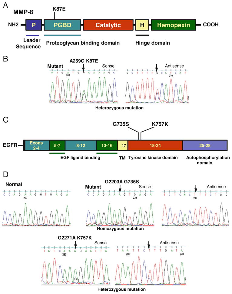
Fig. 1.
Detection of MMP8 and EGFR mutations. a Schematic diagram of domains of MMP8 protein showing a single nucleotide polymorphism (K87E) identified in thyroid cancer. The MMP8 gene is located on chromosome 11q22.3 contains 10 exons and intervening sequences. b The sequencing results were shown with a representative sense and antisense sequence profile of a single nucleotide polymorphisms (A259G) found in exon 2 of MMP8 gene. c Schematic diagram of EGFR showing a mutation (G735S) and a single nucleotide polymorphism (K757K) identified in thyroid cancer. The EGFR gene is located on chromosome 7p11.2 contains 28 exons and intervening sequences. d The sequencing results were shown with sense and antisense sequence profiles of a mutation (G2203A) and a single nucleotide polymorphism (G2271A) found in exon 19 of EGFR gene. Arrow indicates mutated nucleotide. The nucleotide and amino acid alterations are indicated above the arrow. Nucleotide numbers refers to the position within coding sequence, where position 1 corresponds to the first position of the initiation codon. All the samples were sequenced in two repeated examinations with independent PCR by forward and reverse primers

