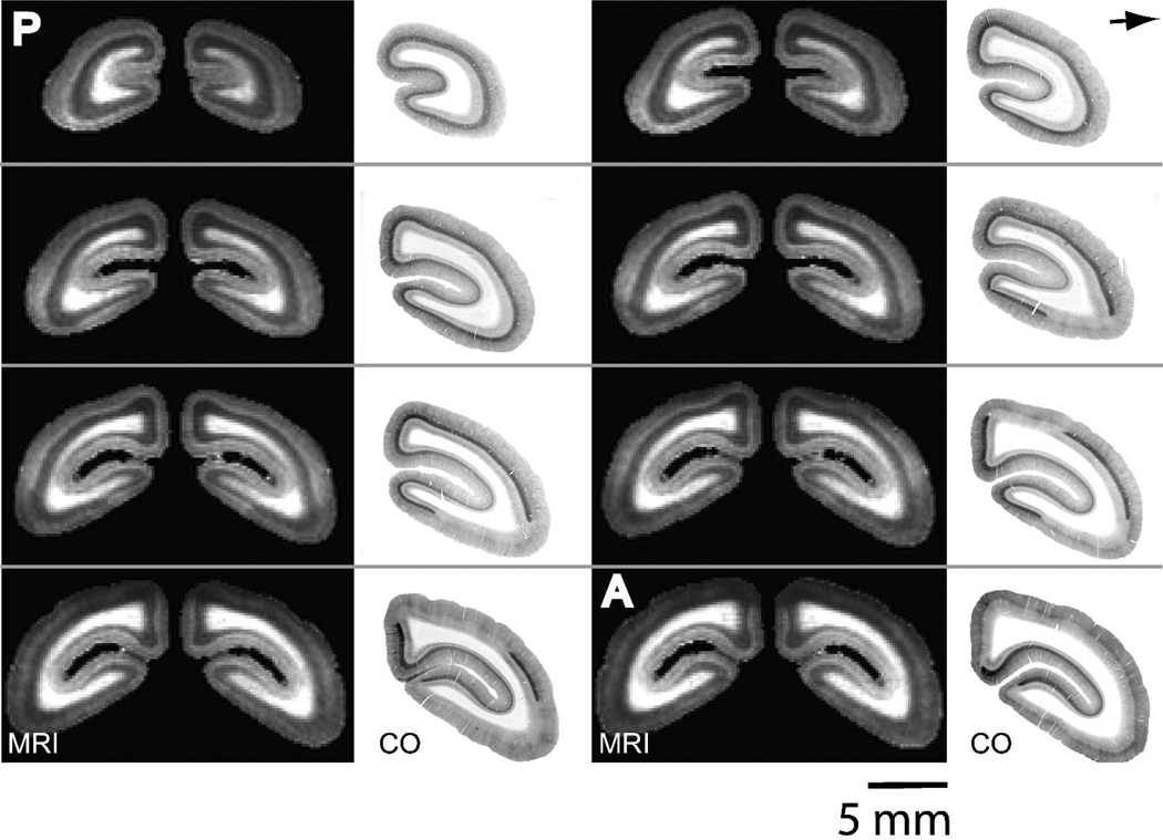
Figure 3.
The extent of V1 is identically identified across the occipital cortex with manganese-enhanced MRI and CO staining. 167 µm in vivo T1-weighted MRI slices and corresponding 40 µm thick CO sections through the occipital cortex. The area of T1 enhancement in the MRI slices corresponds well to V1 as defined by the dark staining in layer IV in the CO sections. The MRI slices are from a 3D MRI in a single marmoset. Slices begin 2.0 mm anterior to the occipital pole and continue in the anterior direction with 0.84 mm spacing. The CO slices are from four marmosets. (P = posterior, A = anterior).

