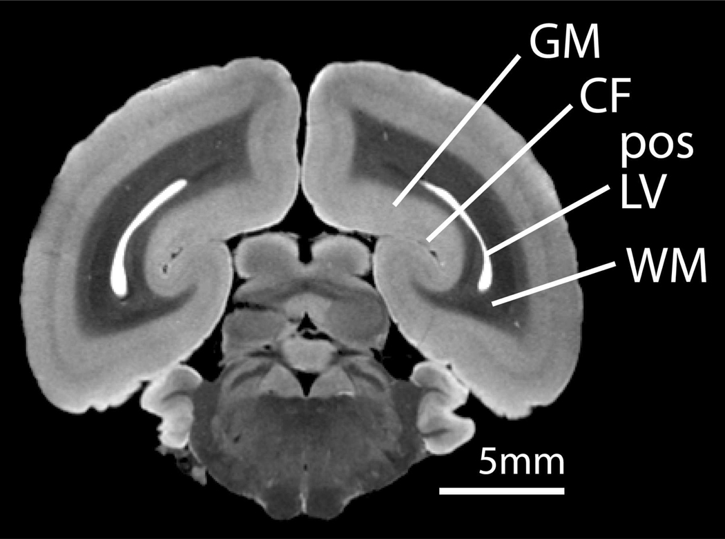
Figure 5.
An ex vivo coronal MR image of a fixed marmoset brain showing the position of the lateral ventricles in the occipital lobe. Gray matter surrounding the calcarine fissure in the occipital cortex is supplied with Mn2+ ions via a CSF-brain uptake route. (GM = gray matter, CF = calcarine fissure, pos LV = posterior horn of the lateral ventricle, WM = white matter).

