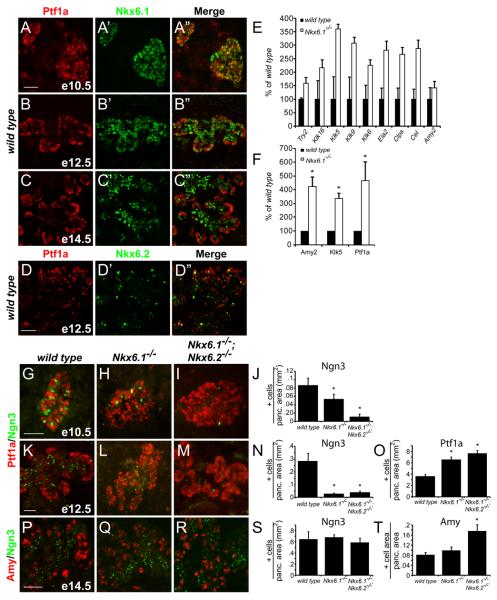
Figure 1. Endocrine to acinar fate conversion in Nkx6-deficient embryos.
(A-D) Immunofluorescence for Ptf1a and Nkx6.1 or Nkx6.2 in the embryonic mouse pancreas. At e12.5, Nkx6.1 and Nkx6.2 become excluded from the Ptf1a+ tip domain. (E-F) Quantification of mRNA levels for acinar markers by microarray (E) and qRT-PCR (F) in e15.5 pancreas. Nkx6.1-deficiency results in increased expression of acinar-specific genes. (G-I,K-M,P-R) Immunofluorescence staining reveals expansion of the Ptf1a+ domain into the trunk region in Nkx6.1−/− and Nkx6.1−/−;Nkx6.2−/− pancreata at e12.5. Concomitantly, Ngn3+ cells are reduced. At e14.5, Nkx6-deficient embryos show increased numbers of acinar cells. (J,N,O,S,T) Quantification of marker+ cell numbers relative to total pancreatic area. Scale bar=50 μm; Try, trypsin; Klk, kallikrein; Ela, elastase; Clps, co-lipase; Cel, carboxylester-lipase; Amy, amylase.

