
Fig. 5.
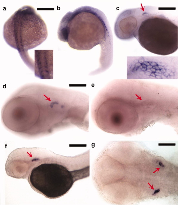
Expression pattern of zCyr61-c5 detected by in situ hybridization. a–g: Photographs of zebrafish embryos staged at 24 (a,b), 48 (c), 55 (d,e), and 70 (f,g) hours postfertilization. Expression is observed in the somites (a,b) and otic vesicle (c,d,f,g, arrows). A sense probe (e), used as a negative control, demonstrates the specificity of zCyr61-c5 expression in the otic vesicle. Inserts in a and c show higher magnification views of somites (a) and otic vesicle (c). Scale bars = 250 μm in a,b, 200 μm in c,f, and 100 μm in d,e,g.